Age-Related Macular Degeneration (AMD): Dry Forms Including Geographic Atrophy
Dry age-related macular degeneration (dry AMD) is the most common form of macular degeneration and a leading cause of vision loss in adults over age 50. Dry AMD affects the macula, the central portion of the retina responsible for sharp vision needed for reading, driving, and recognizing faces.
Dry AMD usually progresses slowly over time. In some patients, it can advance to a more severe stage called geographic atrophy (GA), where areas of retinal tissue gradually deteriorate and stop functioning.
Although dry AMD does not usually cause complete blindness, it can significantly affect central vision and quality of life.
What Causes Dry AMD?
Dry AMD develops as the retina ages. Tiny yellow deposits called drusen build up underneath the retina and may interfere with normal retinal function. Over time, retinal cells can become damaged and die, leading to thinning of the macula.
The exact cause is not fully understood, but several factors increase the risk of dry AMD:
- Age over 50
- Smoking
- Family history of AMD
- High blood pressure
- High cholesterol
- Obesity
- Poor diet
- Excessive sun exposure
Smoking is one of the most important controllable risk factors and can greatly increase the risk of advanced AMD.
What Is Geographic Atrophy?
Geographic atrophy is an advanced form of dry AMD. In GA, patches of retinal tissue gradually degenerate, creating areas where vision no longer functions properly.
These areas of atrophy can slowly enlarge over time and may eventually involve the center of the macula, causing significant vision loss.
Unlike wet AMD, geographic atrophy does not involve bleeding or leaking blood vessels. However, it can still lead to severe central vision impairment.
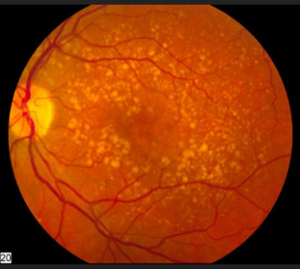
The photograph above shows many drusen throughout the macula representing dry age-related macular degeneration.
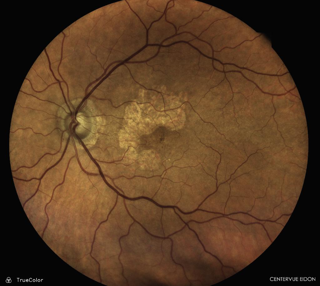
The photograph above has a lighter-colored patch that represents atrophy of the retina near the fovea. This area is an area of geographic atrophy.
Symptoms of Dry AMD and Geographic Atrophy
Symptoms often develop gradually and may worsen slowly over several years.
Common symptoms include:
- Blurred central vision
- Difficulty reading
- Trouble recognizing faces
- Needing brighter light
- Reduced contrast sensitivity
- Missing or dark spots in central vision
- Difficulty adapting to dim lighting
In early dry AMD, patients may not notice any symptoms at all.
As geographic atrophy progresses, blind spots in central vision can become larger and more noticeable.
Peripheral (side) vision is usually preserved.
How Is Dry AMD Diagnosed?
A retinal specialist can diagnose dry AMD during a dilated eye examination. Several imaging tests help determine the severity of disease and monitor progression.
Optical Coherence Tomography (OCT)
OCT creates detailed cross-sectional images of the retina and can show drusen, retinal thinning, and areas of geographic atrophy.
Fundus Photography
Retinal photographs document the appearance and progression of drusen and atrophic changes.
Fundus Autofluorescence
This imaging test highlights areas of retinal damage and can help measure the size and growth of geographic atrophy.
Amsler Grid
Patients may use an Amsler grid at home to monitor for changes in central vision or distortion.
Treatment for Dry AMD and Geographic Atrophy
Lifestyle and Nutritional Support
Although dry AMD cannot currently be cured, several steps may help slow progression:
- Stop smoking
- Eat a healthy diet rich in leafy greens and fish
- Exercise regularly
- Control blood pressure and cholesterol
- Protect eyes from excessive sunlight
Certain patients with intermediate AMD may benefit from AREDS2 vitamin supplements, which have been shown to reduce the risk of progression to advanced AMD.
Your retina specialist can help determine whether AREDS2 supplements are appropriate for you.
Valeda Light Therapy is also a recently-approved treatment for some forms of age-related macular degeneration.
Treatments for Geographic Atrophy
New medications have recently become available to help slow the progression of geographic atrophy.
These treatments include:
- Pegcetacoplan
- Avacincaptad pegol
These medications are given as injections into the eye and are designed to slow the enlargement of atrophic areas. While they do not restore lost vision, they may help preserve vision longer by slowing disease progression.
Not every patient with GA is a candidate for treatment, and treatment plans are individualized based on the location and severity of atrophy.
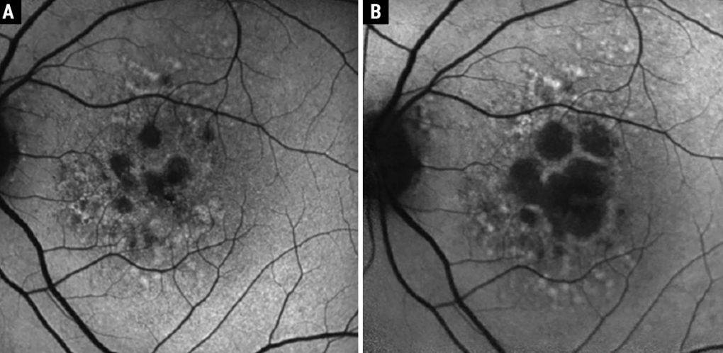
The photo below shows an untreated patient with geographic atrophy that had progression of disease from photo A to photo B. The goal of treatment is to slow this progression as much as possible.
Living With Dry AMD
Many patients with dry AMD continue normal daily activities for years. Helpful strategies include:
- Using brighter lighting
- Enlarging text on digital devices
- Using magnifiers
- Scheduling regular retina examinations
- Monitoring vision changes at home
Low vision specialists can provide tools and training to help maximize remaining vision.
Frequently Asked Questions (FAQ)
Does dry AMD always become wet AMD?
No. Many patients with dry AMD never develop wet AMD. However, patients with dry AMD do have an increased risk of developing wet AMD over time.
Can geographic atrophy be reversed?
Currently, there is no way to reverse geographic atrophy or restore damaged retinal tissue. New treatments are designed to slow progression rather than cure the disease.
Are vitamin supplements helpful?
Certain patients with intermediate AMD may benefit from AREDS2 supplements. These vitamins are not recommended for everyone, so patients should discuss supplementation with their retina specialist.