Branch Retinal Vein Occlusion (BRVO)
Branch retinal vein occlusion (BRVO) is a common retinal vascular condition that occurs when one of the small veins in the retina becomes blocked. The retina is the light-sensitive tissue lining the back of the eye, and it is responsible for sending visual signals to the brain.
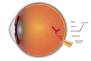
When a retinal vein becomes blocked, blood and fluid can leak into the retina, leading to swelling, bleeding, and blurred vision. BRVO usually affects only one eye and most commonly occurs in adults over age 50.
The severity of BRVO can vary widely. Some patients have only mild vision changes, while others may experience more significant vision loss, especially if the center of the retina (the macula) becomes swollen.
What Causes BRVO?
BRVO occurs when a retinal vein becomes compressed or blocked, often at a location where a retinal artery crosses over the vein. This blockage interferes with normal blood flow and causes pressure to build up inside the retinal vessels.
The most common risk factors include:
- High blood pressure
- Diabetes
- High cholesterol
- Smoking
- Cardiovascular disease
- Glaucoma
- Aging
Many patients with BRVO have underlying vascular conditions that affect blood circulation throughout the body.
Symptoms of BRVO
Symptoms can appear suddenly or gradually depending on the severity of the blockage.
Common symptoms include:
- Blurred vision
- Distorted central vision
- Dark spots or areas of missing vision
- Difficulty reading
- Sudden painless vision loss in one eye
Some patients may notice only mild blurry vision, while others experience more severe changes if swelling develops in the macula.
Peripheral vision is often less affected than central vision.
How Is BRVO Diagnosed?
A retinal specialist can usually diagnose BRVO during a dilated eye examination. The retina may show bleeding, swollen blood vessels, and retinal swelling.
Several imaging tests help confirm the diagnosis and guide treatment.
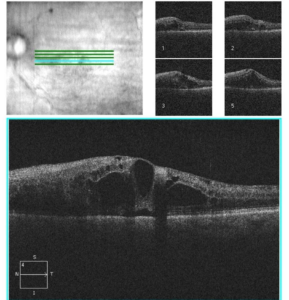
Optical Coherence Tomography (OCT)
OCT creates detailed cross-sectional images of the retina and helps detect swelling in the macula, called macular edema.
Below is an OCT scan showing intraretinal fluid consistent with macular edema.
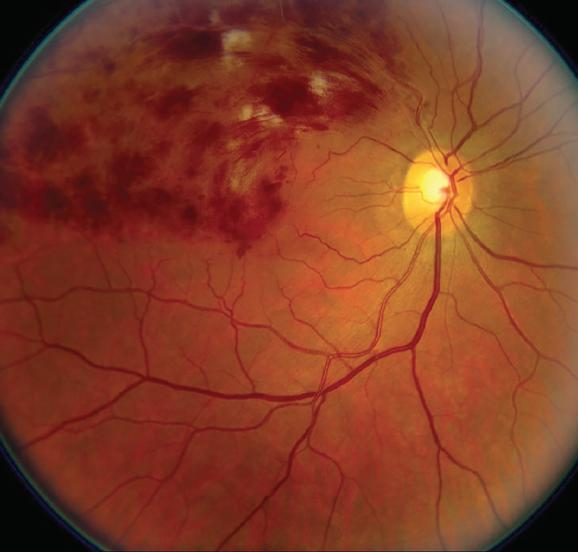
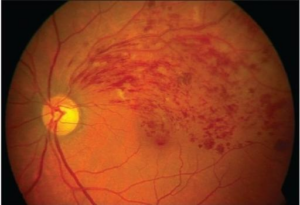
Fundus Photography
Retinal photographs help document bleeding and monitor changes over time.
The photograph below represents a color fundus photo of an eye with a branch retinal vein occlusion showing scattered retinal hemorrhages in the superior macula.
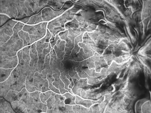
Fluorescein Angiography
This test uses a fluorescent dye injected into a vein in the arm to evaluate blood flow in the retina and identify areas of leakage or poor circulation.
The image below is a fluorescein angiogram showing blockage of dye from scattered retinal hemorrhages.
Complications of BRVO
The most common complication of BRVO is macular edema, which occurs when fluid leaks into the central retina and causes swelling.
Other possible complications include:
- Retinal ischemia (poor blood flow)
- Abnormal blood vessel growth (neovascularization)
- Vitreous hemorrhage
- Elevated eye pressure or glaucoma
Careful monitoring is important because some complications can threaten vision if untreated.
Treatment for BRVO
Treatment depends on the severity of the blockage and whether complications develop.
Anti-VEGF Injections
The most common treatment for macular edema from BRVO is anti-VEGF medication injected into the eye.
These medications help reduce swelling and leakage.
Common anti-VEGF medications include:
- Aflibercept
- Ranibizumab
- Bevacizumab
- Faricimab
Many patients require a series of injections over time.
Steroid Injections or Implants
In some cases, corticosteroid medications may help reduce retinal swelling.
Laser Treatment
Laser treatment may occasionally be used to treat areas of poor retinal circulation or abnormal blood vessel growth.
Managing Systemic Health
Controlling blood pressure, cholesterol, diabetes, and other vascular risk factors is extremely important.
What Is the Prognosis?
The visual outlook for BRVO varies depending on:
- The location of the blockage
- The amount of retinal swelling
- The level of retinal blood flow
- How quickly treatment begins
Many patients experience improvement in vision with modern treatment, especially when macular edema is treated early.
Some patients may continue to have mild blurry or distorted vision even after swelling improves.
Living With BRVO
Patients with BRVO should:
- Keep regular retina appointments
- Monitor for sudden changes in vision
- Maintain healthy blood pressure and cholesterol levels
- Avoid smoking
- Follow treatment recommendations carefully
Long-term follow-up is important because recurrent swelling can occur.
Frequently Asked Questions (FAQ)
Is BRVO a stroke in the eye?
BRVO is sometimes described as an “eye stroke” because it involves blockage of a retinal blood vessel. However, it is different from a brain stroke. Patients with BRVO should still discuss cardiovascular risk factors with their primary care physician.
Can BRVO go away on its own?
Some mild cases improve without treatment, but many patients require injections or monitoring for complications such as macular edema.
Will my vision return to normal?
Many patients improve with treatment, but the amount of recovery varies. Early diagnosis and treatment often lead to better outcomes.