Choroidal Nevus
A choroidal nevus is a benign (non-cancerous) pigmented spot inside the eye, similar to a freckle or mole on the skin. It develops in the choroid, the layer of blood vessels underneath the retina that helps nourish the eye.
Choroidal nevi are common and are often discovered during a routine dilated eye examination. Most choroidal nevi do not cause symptoms and remain stable throughout life. However, because a small percentage can develop into a cancerous tumor called choroidal melanoma, regular monitoring by an eye specialist is important.
What Causes a Choroidal Nevus?
A choroidal nevus develops when pigment-producing cells called melanocytes accumulate within the choroid.
The exact cause is not fully understood, but choroidal nevi are thought to develop similarly to freckles or moles elsewhere in the body.
Most choroidal nevi:
- Are benign
- Grow very slowly or not at all
- Remain stable over time
Choroidal nevi are usually found in adults and may become more noticeable with age.
Symptoms of Choroidal Nevus
Most patients with a choroidal nevus have no symptoms.
When symptoms do occur, they may include:
- Blurred vision
- Distorted vision
- Flashes or floaters
- A shadow in vision
Symptoms are more likely if the nevus develops fluid leakage or is located near the macula, the center of the retina responsible for sharp vision.
Many patients learn they have a nevus only after a routine eye examination.
How Is a Choroidal Nevus Diagnosed?
A retinal specialist or ophthalmologist can usually diagnose a choroidal nevus during a dilated eye examination.
Several imaging tests help document the nevus and monitor for changes over time.
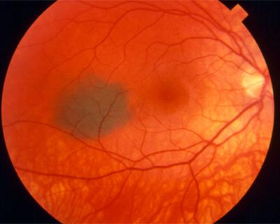
Fundus Photography
Retinal photographs provide a detailed record of the size, color, and appearance of the nevus.
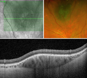
Optical Coherence Tomography (OCT)
OCT creates cross-sectional images of the retina and can detect fluid or swelling associated with the nevus.
Ultrasound Imaging
Ultrasound may be used to measure the thickness and internal structure of the lesion.
Fundus Autofluorescence
This imaging test can help detect orange pigment or retinal changes that may suggest increased risk of growth.
Can a Choroidal Nevus Become Cancer?
Most choroidal nevi never become cancerous. However, a small percentage can transform into choroidal melanoma, a type of eye cancer.
Retina specialists monitor for warning signs that may increase the risk of transformation.
Risk factors include:
- Increased thickness
- Fluid beneath the retina
- Orange pigment
- Growth over time
- Symptoms such as blurred vision
- Location near the optic nerve
Because of this small risk, regular follow-up examinations are important.
How Often Should a Choroidal Nevus Be Monitored?
Monitoring schedules vary depending on the appearance of the nevus and whether risk factors are present.
Typical follow-up may include:
- Every 4–6 months initially
- Annual examinations if stable
- More frequent imaging if suspicious features develop
Many nevi remain unchanged for years.

Treatment for Choroidal Nevus
Most choroidal nevi do not require treatment.
Instead, treatment focuses on:
- Careful monitoring
- Retinal imaging
- Detecting growth or suspicious changes early
If a nevus develops features concerning for melanoma, referral to an ocular oncology specialist may be recommended.
When Is Treatment Needed?
Treatment is generally considered only if:
- The lesion shows growth
- Suspicious features appear
- Vision-threatening fluid develops
- Melanoma is suspected
Treatment for choroidal melanoma may include radiation therapy, laser treatment, or surgery.
What Is the Prognosis?
The outlook for patients with a choroidal nevus is usually excellent.
Most nevi:
- Remain benign
- Cause no symptoms
- Require only periodic monitoring
Early detection of suspicious changes allows prompt treatment if necessary.
Living With a Choroidal Nevus
Patients with a choroidal nevus should:
- Keep regular eye appointments
- Report new visual symptoms promptly
- Maintain routine retinal imaging
- Understand that most nevi are harmless
Learning that you have a “spot” inside the eye can be concerning, but the vast majority of choroidal nevi never become dangerous.
Frequently Asked Questions (FAQ)
Is a choroidal nevus cancer?
No. A choroidal nevus is usually a benign freckle or mole inside the eye. However, because a small percentage can develop into melanoma, regular monitoring is important.
Can a choroidal nevus affect vision?
Most nevi do not affect vision. Vision changes are more likely if the nevus leaks fluid or is located near the macula.
Will I need treatment?
Most patients do not need treatment and only require periodic monitoring with retinal imaging and examinations.