Sickle Cell Retinopathy
Sickle cell retinopathy is an eye condition that occurs when sickle cell disease affects the blood vessels of the retina, the light-sensitive tissue at the back of the eye. Changes in these small retinal blood vessels can reduce blood flow, cause abnormal new blood vessel growth, and, in severe cases, lead to vision-threatening complications.
Many people with sickle cell retinopathy have no symptoms in the early stages. Because retinal changes can develop silently, regular dilated eye examinations are an important part of care for anyone with sickle cell disease or certain forms of sickle cell trait.
With early detection and appropriate treatment, many patients can maintain excellent vision throughout their lives.
What Is Sickle Cell Disease?
Sickle cell disease is an inherited blood disorder that affects hemoglobin, the protein inside red blood cells that carries oxygen throughout the body.
Normally, red blood cells are round and flexible, allowing them to move easily through blood vessels. In sickle cell disease, red blood cells can become rigid and crescent-shaped (“sickle-shaped”), especially under conditions of stress or low oxygen.
These abnormal cells can block small blood vessels, reducing blood flow to tissues throughout the body—including the retina.
How Does Sickle Cell Disease Affect the Eye?
The retina requires a constant supply of oxygen and nutrients. When sickled red blood cells obstruct retinal blood vessels, areas of the retina may become deprived of oxygen.
This can lead to:
- Retinal ischemia (poor blood flow)
- Closure of small retinal vessels
- Abnormal blood vessel growth (neovascularization)
- Bleeding into the vitreous gel
- Scar tissue formation
- Retinal detachment
Not everyone with sickle cell disease develops significant retinal complications, but regular screening remains essential.
Who Is at Risk?
Sickle cell retinopathy can occur in individuals with several hemoglobin disorders, including:
- Sickle cell disease (HbSS)
- Hemoglobin SC disease (HbSC)
- Sickle beta-thalassemia
Patients with HbSC disease often develop more severe proliferative retinal disease than those with HbSS disease.
Risk factors for developing retinal complications include:
- Increasing age
- Longer duration of disease
- Certain sickle cell genotypes
- Poor access to routine eye examinations
Symptoms of Sickle Cell Retinopathy
Many patients have no symptoms during the early stages of the disease.
When symptoms occur, they may include:
- Floaters
- Blurred vision
- Sudden vision loss
- Dark spots in vision
- Distorted vision
- Loss of part of the visual field
Sudden vision changes may indicate vitreous hemorrhage or retinal detachment and require urgent evaluation.
Stages of Sickle Cell Retinopathy
Doctors often classify sickle cell retinopathy using the Goldberg classification system.
Stage I: Peripheral Arteriolar Occlusions
Small retinal blood vessels become blocked.
Stage II: Arteriovenous Anastomoses
New connections form between blood vessels in response to poor circulation.
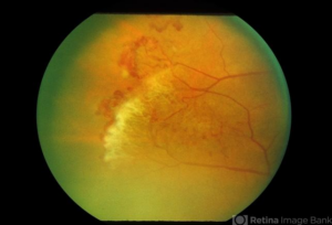
Stage III: Neovascularization
Abnormal new blood vessels develop at the border between healthy and ischemic retina.
These fragile blood vessels often have a characteristic appearance called “sea fan” neovascularization.
Stage IV: Vitreous Hemorrhage
The abnormal vessels can bleed into the vitreous cavity.
Stage V: Retinal Detachment
Scar tissue and traction can pull the retina away from the back wall of the eye.
How Is Sickle Cell Retinopathy Diagnosed?
A retina specialist can diagnose sickle cell retinopathy through a comprehensive examination and specialized imaging.
Dilated Retinal Examination
Your doctor examines the retina for:
- Areas of poor circulation
- Sea fan neovascularization
- Vitreous hemorrhage
- Retinal tears
- Retinal detachment
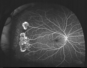
Fluorescein Angiography
This imaging test uses a special dye to evaluate retinal blood flow and identify:
- Areas of nonperfusion
- Abnormal new blood vessels
- Leakage from neovascularization
Optical Coherence Tomography (OCT)
OCT provides detailed cross-sectional images of the retina and can identify:
- Macular thinning
- Macular ischemia
- Structural changes affecting central vision
Wide-Field Retinal Imaging
Wide-field photography can document peripheral retinal abnormalities and monitor disease progression over time.
How Is Sickle Cell Retinopathy Treated?
Treatment depends on the stage of the disease and the presence of complications.
Observation
Early stages without neovascularization may only require monitoring with regular examinations.
Laser Photocoagulation
Laser treatment may be recommended for proliferative disease.
The laser helps:
- Reduce oxygen demand in ischemic retina
- Cause regression of abnormal blood vessels
- Lower the risk of vitreous hemorrhage
Anti-VEGF Injections
In selected cases, anti-VEGF medications may help reduce neovascularization and assist in managing complications.
Vitrectomy Surgery
Surgery may be necessary for patients with:
- Non-clearing vitreous hemorrhage
- Tractional retinal detachment
- Combined retinal detachments
During vitrectomy, the vitreous gel and scar tissue are removed to restore or preserve vision.
What Is the Prognosis?
The outlook for patients with sickle cell retinopathy is generally favorable when the disease is detected early.
Many patients:
- Maintain excellent vision
- Require only observation
- Never develop advanced complications
However, untreated proliferative disease can lead to permanent vision loss from vitreous hemorrhage or retinal detachment.
Regular screening remains the best way to prevent serious complications.
Living With Sickle Cell Retinopathy
If you have sickle cell disease or a related hemoglobin disorder:
- Schedule regular dilated retinal examinations.
- Follow recommendations from your hematologist and retina specialist.
- Seek urgent evaluation for new floaters or sudden vision changes.
- Maintain good overall health and hydration.
- Inform your eye doctor about your sickle cell diagnosis.
Coordinated care between your medical team and eye specialists helps optimize both systemic and ocular health.
Frequently Asked Questions (FAQ)
Can sickle cell disease cause blindness?
Yes, severe retinal complications can lead to vision loss. Fortunately, regular eye examinations and timely treatment greatly reduce this risk.
If I have sickle cell disease but no symptoms, do I still need eye exams?
Yes. Early sickle cell retinopathy often causes no symptoms. Routine screening allows problems to be detected before vision is affected.
What is "sea fan" neovascularization?
Sea fan neovascularization refers to abnormal new blood vessels that develop in proliferative sickle cell retinopathy. Their branching appearance resembles a sea fan coral and can lead to bleeding if left untreated.