
Posterior Vitreous Detachment (PVD)
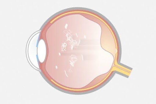
A posterior vitreous detachment (PVD) is a common age-related change that occurs when the vitreous gel inside the eye separates from the retina. Although a PVD is usually a normal part of aging and often does not threaten vision, it can sometimes lead to retinal tears or retinal detachment.
Because the symptoms of a PVD can overlap with those of more serious retinal conditions, any sudden onset of flashes, floaters, or changes in vision should be evaluated promptly by an eye care professional.
What Is the Vitreous?
The vitreous is a clear, gel-like substance that fills the inside of the eye and helps maintain its shape. In younger individuals, the vitreous has a firm consistency and is attached to the retina in several locations.
As we age:
- The vitreous gradually becomes more liquid.
- The gel shrinks and collapses.
- The vitreous begins to pull away from the retina.
- Eventually, it separates from the retinal surface.
This separation is called a posterior vitreous detachment.
What Causes a Posterior Vitreous Detachment?
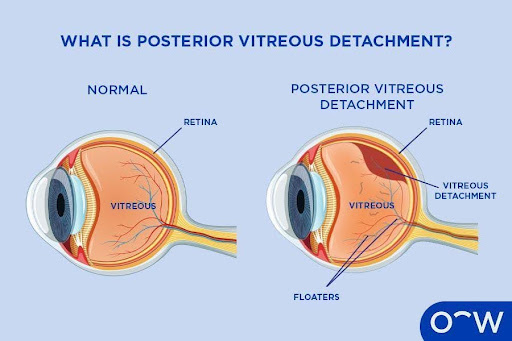
PVD is most often related to normal aging.
Risk factors include:
- Age over 50 years
- Nearsightedness (myopia)
- Previous cataract surgery
- Eye trauma
- Inflammation inside the eye (uveitis)
- A history of PVD in the other eye
A PVD is very common and becomes increasingly likely with advancing age.
Symptoms of a PVD
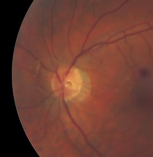
The symptoms of a PVD often develop suddenly.
Common symptoms include:
Floaters
Floaters may appear as:
- Small dots
- Cobwebs
- Threads
- Rings
- Shadows drifting through the field of vision
Many patients notice a large ring-shaped floater, sometimes called a Weiss ring, which occurs when the vitreous separates from the optic nerve.
Flashes of Light
Flashes occur when the vitreous pulls on the retina during the separation process.
Patients often describe them as:
- Brief streaks of light
- Lightning-like flashes
- Flickers in the peripheral vision
Blurred Vision
Temporary blurring can occur if floaters pass through the line of sight.
No Symptoms
Some people develop a PVD without noticing any symptoms.
Why Is a PVD Important?
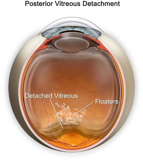
Most PVDs do not cause serious complications.
However, as the vitreous separates from the retina, traction can occasionally create a retinal tear.
If untreated, a retinal tear can allow fluid to pass beneath the retina and lead to a retinal detachment.
Approximately 10–15% of patients with an acute symptomatic PVD may have an associated retinal tear at the time of diagnosis.
Warning Signs of a Retinal Tear or Detachment
Seek urgent evaluation if you experience:
- A sudden increase in floaters
- New flashes of light
- A curtain or shadow coming across your vision
- Loss of peripheral vision
- Sudden decrease in vision
These symptoms may indicate a retinal tear or retinal detachment and should not be ignored.
How Is a PVD Diagnosed?
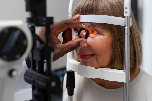
A retinal specialist can diagnose a PVD through a comprehensive dilated eye examination.
Dilated Retinal Examination
Special lenses are used to examine the retina for:
- Posterior vitreous separation
- Retinal tears
- Retinal holes
- Retinal detachment
- Vitreous hemorrhage
Optical Coherence Tomography (OCT)
OCT may help evaluate the vitreomacular interface and identify associated macular conditions.
Ultrasound Imaging
Ultrasound may be used if the retina cannot be visualized clearly because of vitreous hemorrhage or cataracts.
Treatment for Posterior Vitreous Detachment
Observation
Most PVDs require no treatment.
Over time:
- Floaters often become less noticeable.
- The brain adapts to their presence.
- Flashes usually decrease and resolve.
Treatment of Retinal Tears
If a retinal tear is found, treatment may include:
- Laser retinopexy
- Cryotherapy
These procedures help seal the retina and reduce the risk of retinal detachment.
Treatment of Retinal Detachment
If retinal detachment develops, surgery may be necessary.
Treatment options include:
- Pneumatic retinopexy
- Scleral buckle surgery
- Pars plana vitrectomy
Treatment for Persistent Floaters
In selected patients with severe, visually disabling floaters, vitrectomy surgery may be considered after careful discussion of risks and benefits.
What Is the Prognosis?
The outlook for most patients with PVD is excellent.
Most individuals:
- Do not develop retinal complications.
- Maintain normal vision.
- Experience improvement in symptoms over time.
The greatest risk period for retinal tears is during the early stages of vitreous separation, making prompt evaluation of new symptoms essential.
Living With a PVD
If you have been diagnosed with a PVD:
- Attend all recommended follow-up appointments.
- Monitor for new flashes or floaters.
- Seek immediate care if you notice a curtain or shadow in your vision.
- Protect your eyes from trauma.
- Inform your eye doctor if similar symptoms develop in the other eye.
A PVD in one eye increases the likelihood of developing a PVD in the fellow eye over time.
Frequently Asked Questions (FAQ)
Is a posterior vitreous detachment the same as a retinal detachment?
No. A PVD occurs when the vitreous separates from the retina. A retinal detachment occurs when the retina itself pulls away from the back wall of the eye and is a vision-threatening emergency.
Will my floaters go away?
Floaters often become less noticeable over weeks to months as they settle and the brain adapts to them. Some floaters may persist long term.
Should I be worried about flashes and floaters?
Any sudden onset of flashes, floaters, or changes in vision should be evaluated promptly because these symptoms can indicate a retinal tear or retinal detachment.
More Information
For additional patient information, visit the American Society of Retina Specialists Posterior Vitreous Detachment Page