Age-Related Macular Degeneration (AMD): Wet Forms Including Macular Neovascularization
Wet age-related macular degeneration (wet AMD) is a serious form of macular degeneration that can cause rapid central vision loss if left untreated. Wet AMD develops when abnormal blood vessels grow underneath the retina and macula, a process called macular neovascularization (MNV) or choroidal neovascularization (CNV).
These abnormal blood vessels are fragile and may leak fluid, blood, or scar tissue beneath the retina. This can damage the macula and interfere with the sharp central vision needed for reading, driving, and recognizing faces.
Although wet AMD accounts for only about 10–20% of AMD cases, it causes the majority of severe vision loss related to macular degeneration.
What Causes Wet AMD?
Wet AMD usually develops from underlying dry AMD. As retinal tissue becomes damaged with age, the eye may produce signals that stimulate the growth of abnormal blood vessels beneath the retina.
These vessels can leak and bleed, leading to swelling and scarring of the macula.
Several factors increase the risk of wet AMD:
- Age over 50
- Smoking
- Family history of AMD
- High blood pressure
- High cholesterol
- Cardiovascular disease
- Obesity
- Previous dry AMD
Smoking is one of the strongest risk factors and significantly increases the chance of developing advanced AMD.
What Is Macular Neovascularization (MNV)?
Macular neovascularization refers to the abnormal growth of blood vessels beneath the macula. These vessels originate from deeper layers beneath the retina and grow into areas where they should not normally exist.
MNV can cause:
- Fluid accumulation
- Bleeding
- Retinal swelling
- Scar tissue formation
- Permanent central vision damage
Prompt diagnosis and treatment are extremely important because untreated MNV can lead to irreversible vision loss.
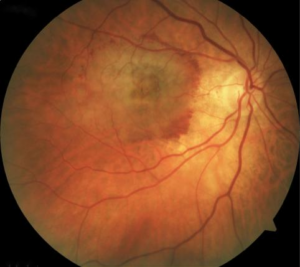
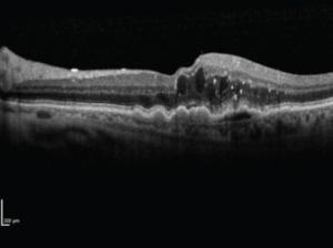
Below are fundus photos and an OCT scan representing changes including drusen, changes in the retinal pigment epithelium, and fluid accumulation that is caused by age-related macular degeneration with macular neovascularization.
Symptoms of Wet AMD
Wet AMD symptoms often develop suddenly and may worsen quickly.
Common symptoms include:
- Blurred central vision
- Distorted vision (straight lines appear bent or wavy)
- Dark or missing spots in central vision
- Difficulty reading
- Trouble recognizing faces
- Rapid decline in visual clarity
- Colors appearing faded
Many patients first notice distortion while reading or looking at door frames, window blinds, or tile lines.
Peripheral (side) vision is usually preserved.
How Is Wet AMD Diagnosed?
A retinal specialist can diagnose wet AMD during a dilated eye examination. Specialized imaging tests help confirm the presence of fluid or abnormal blood vessels.
Optical Coherence Tomography (OCT)
OCT is the most important imaging test for wet AMD. It creates detailed cross-sectional images of the retina and can detect:
- Fluid beneath the retina
- Retinal swelling
- Bleeding
- Pigment epithelial detachments
OCT Angiography (OCTA)
OCTA can visualize abnormal blood vessels without the need for dye injections.
Fluorescein Angiography
This test uses a fluorescent dye injected into a vein in the arm to identify leaking blood vessels beneath the retina.
Fundus Photography
Retinal photographs help document bleeding, swelling, and long-term disease progression.
Treatment for Wet AMD
The primary treatment for wet AMD is anti-VEGF therapy. VEGF (vascular endothelial growth factor) is a protein that promotes abnormal blood vessel growth and leakage.
Anti-VEGF medications block this process and help reduce fluid and bleeding.
Common anti-VEGF medications include:
- Aflibercept
- Ranibizumab
- Bevacizumab
- Faricimab
These medications are given through injections into the eye during a sterile office procedure.
How Often Are Injections Needed?
Treatment schedules vary from person to person. Some patients require monthly injections initially, while others may eventually transition to less frequent treatment intervals.
Regular monitoring with OCT imaging helps guide treatment decisions.
Can Vision Improve?
Many patients experience stabilization or improvement in vision with treatment, especially when therapy begins early. However, untreated wet AMD can lead to permanent scarring and irreversible vision loss.
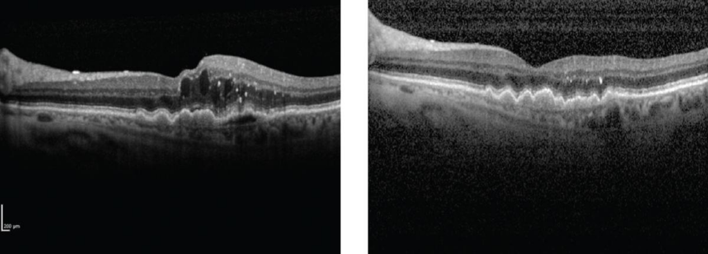
Below are OCT scans before and after treatment with anti-VEGF therapy. These show how intravitreal injections can be an effective way to treat the pathology caused by wet AMD.
Living With Wet AMD
Wet AMD often requires ongoing monitoring and long-term treatment. Helpful strategies include:
- Keeping scheduled retina appointments
- Monitoring vision changes at home
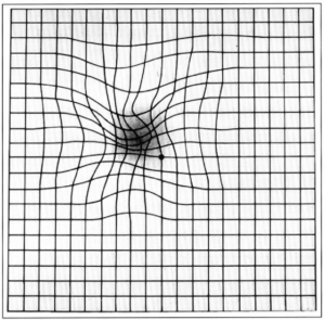
- Using an Amsler grid regularly
- Reporting sudden changes in vision immediately
- Maintaining a healthy lifestyle
- Avoiding smoking
Low vision tools and magnification devices may also help improve daily functioning.
Amsler grid monitoring at home can be a useful monitoring tool. Below is a representation of amsler grid changes that a patient with new visual distortion from macular degeneration may see.
Frequently Asked Questions (FAQ)
Is wet AMD curable?
Wet AMD cannot currently be cured, but modern treatments can often slow progression, stabilize vision, and sometimes improve vision.
Are eye injections painful?
Most patients tolerate injections very well. The eye is numbed beforehand, and patients usually feel pressure rather than pain.
How quickly should wet AMD be treated?
Treatment should begin as soon as possible after diagnosis. Delays can increase the risk of permanent vision loss.