Central Retinal Vein Occlusion (CRVO)
Central retinal vein occlusion (CRVO) is a serious retinal vascular condition that occurs when the main vein draining blood from the retina becomes blocked. The retina is the light-sensitive tissue at the back of the eye that allows us to see.
When the central retinal vein becomes blocked, blood and fluid can back up inside the retina, leading to swelling, bleeding, and decreased vision. CRVO usually affects one eye and most commonly occurs in adults over age 50.
The severity of CRVO can vary significantly. Some patients experience mild blurry vision, while others may develop severe vision loss due to retinal swelling or poor blood flow.
What Causes CRVO?
CRVO develops when the main retinal vein becomes narrowed or blocked, interfering with normal blood circulation inside the eye.
Several medical conditions increase the risk of CRVO, including:
- High blood pressure
- Diabetes
- High cholesterol
- Smoking
- Cardiovascular disease
- Glaucoma
- Blood clotting disorders
- Aging
Many patients with CRVO have underlying vascular disease that affects circulation throughout the body.
Types of CRVO
Non-Ischemic CRVO
This is the more common and generally milder form of CRVO. Blood flow is reduced but not completely blocked.
Patients may have:
- Mild to moderate blurry vision
- Retinal swelling
- Less severe retinal damage
Some patients maintain relatively good vision with treatment.
Ischemic CRVO
This is the more severe form and occurs when there is significant loss of retinal blood flow.
Patients with ischemic CRVO may experience:
- Severe vision loss
- Extensive retinal bleeding
- Poor oxygen supply to the retina
- Increased risk of complications
Ischemic CRVO can lead to abnormal blood vessel growth and glaucoma if not carefully monitored.
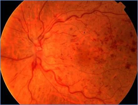
Below is a photograph of a central retinal vein occlusion which shows swollen retinal veins as well as scattered retinal hemorrhages (blood spots).
Symptoms of CRVO
Symptoms usually occur suddenly and affect one eye.
Common symptoms include:
- Blurred vision
- Sudden painless vision loss
- Distorted central vision
- Dark spots or missing areas in vision
- Difficulty reading or recognizing faces
The amount of vision loss depends on the severity of the blockage and whether swelling develops in the macula.
How Is CRVO Diagnosed?
A retinal specialist can usually diagnose CRVO during a dilated eye examination. The retina often shows widespread bleeding, swollen veins, and retinal swelling.
Several imaging tests help determine the severity of disease and guide treatment.
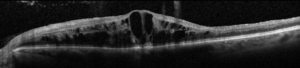
Optical Coherence Tomography (OCT)
OCT creates detailed cross-sectional images of the retina and helps detect macular edema, which is swelling in the center of the retina.
Fluorescein Angiography
This test uses fluorescent dye injected into a vein in the arm to evaluate retinal blood flow and identify areas of poor circulation or leakage.
Fundus Photography
Retinal photographs document retinal hemorrhages and help monitor progression over time.
Complications of CRVO
The most common complication is macular edema, which can significantly reduce central vision.
Other complications may include:
- Retinal ischemia (poor blood flow)
- Neovascularization (abnormal blood vessel growth)
- Vitreous hemorrhage
- Neovascular glaucoma
- Permanent vision loss
Patients with ischemic CRVO require especially close monitoring because complications can develop rapidly.
Treatment for CRVO
Treatment focuses on reducing retinal swelling, preventing complications, and preserving vision.
Anti-VEGF Injections
Anti-VEGF medications are the most common treatment for macular edema caused by CRVO.
These medications help reduce fluid leakage and swelling in the retina.
Common anti-VEGF medications include:
- Aflibercept
- Ranibizumab
- Bevacizumab
- Faricimab
Treatment often requires repeated injections over time.
Steroid Injections or Implants
Steroid medications may also help reduce retinal swelling in selected patients.
Laser Treatment
Laser treatment may be used if abnormal blood vessels develop due to poor retinal circulation.
Managing Systemic Health
Control of blood pressure, diabetes, cholesterol, and other cardiovascular risk factors is extremely important.
What Is the Prognosis?
Visual outcomes vary depending on:
- Whether the CRVO is ischemic or non-ischemic
- The degree of macular edema
- The amount of retinal blood flow
- How quickly treatment begins
Some patients improve substantially with treatment, while others may have permanent vision loss despite therapy.
Regular follow-up is essential because complications can develop even months after diagnosis.
Living With CRVO
Patients with CRVO should:
- Keep regular retina appointments
- Monitor vision changes carefully
- Control blood pressure and diabetes
- Avoid smoking
- Follow injection schedules as recommended
Prompt evaluation of sudden vision changes is very important.
Frequently Asked Questions (FAQ)
Is CRVO considered an eye stroke?
CRVO is often described as an “eye stroke” because it involves blockage of a major retinal blood vessel. Patients with CRVO should discuss cardiovascular risk factors with their primary care physician.
Can CRVO improve over time?
Some patients experience improvement, especially with modern treatments for macular edema. However, recovery varies widely depending on disease severity.
Will I need injections forever?
Some patients require long-term injections, while others may eventually need less frequent treatment. The treatment plan depends on how the retina responds over time.