Choroidal Detachment
A choroidal detachment occurs when fluid or blood collects in the space between the choroid and the sclera inside the eye. The choroid is the layer of blood vessels beneath the retina that helps supply oxygen and nutrients to the eye. The sclera is the white outer wall of the eye.
When this fluid or blood builds up, the choroid separates from the sclera, creating a choroidal detachment. This condition can occur after eye surgery, trauma, inflammation, or very low eye pressure.
The severity of a choroidal detachment can range from mild and temporary to severe and vision-threatening.
What Causes Choroidal Detachment?
Choroidal detachments occur when fluid or blood accumulates within the eye. There are two main types:
Serous Choroidal Detachment
This occurs when clear fluid collects beneath the choroid. Common causes include:
- Low eye pressure after surgery
- Eye inflammation
- Severe hypotony (very low intraocular pressure)
- Certain medications
- Retinal surgery
Hemorrhagic Choroidal Detachment
This occurs when bleeding develops beneath the choroid. Causes may include:
- Eye surgery
- Trauma
- Advanced glaucoma
- Blood-thinning medications
- High blood pressure
Hemorrhagic detachments are often more painful and more serious than serous detachments.
Symptoms of Choroidal Detachment
Symptoms depend on the size and severity of the detachment.
Common symptoms include:
- Blurred vision
- Eye pain or discomfort
- Sensation of pressure in the eye
- Reduced peripheral vision
- Distorted vision
- Redness
- Headache
Some small detachments may cause few or no symptoms and are detected only during an eye examination.
How Is Choroidal Detachment Diagnosed?
A retinal specialist or ophthalmologist can diagnose a choroidal detachment during a dilated eye examination.
Several imaging tests may help confirm the diagnosis and determine severity.
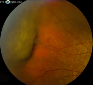
Dilated Eye Examination
The doctor may see elevated, smooth dome-shaped areas inside the eye caused by fluid or blood beneath the choroid.
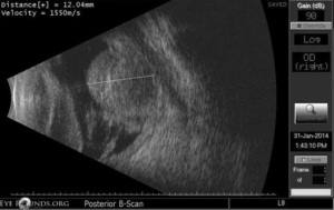
Ultrasound Imaging (B-Scan)
Ultrasound is especially useful when the view into the eye is limited by bleeding or cataracts. It helps visualize the extent of the detachment.
Optical Coherence Tomography (OCT)
OCT may help evaluate associated retinal swelling or other retinal conditions.
Eye Pressure Measurement
Many patients with choroidal detachment have very low eye pressure.
When Does Choroidal Detachment Occur?
Choroidal detachments may occur:
- After glaucoma surgery
- Following retinal surgery
- After eye trauma
- With severe inflammation
- In association with retinal detachment
- After sudden drops in eye pressure
Postoperative choroidal detachments are relatively common after some eye surgeries and often improve with treatment.
Treatment for Choroidal Detachment
Treatment depends on:
- The size of the detachment
- The underlying cause
- Eye pressure
- Whether blood or fluid is present
- The severity of symptoms
Observation
Small serous choroidal detachments may resolve on their own with close monitoring.
Medications
Treatment may include:
- Steroid eye drops
- Oral steroids
- Cycloplegic eye drops
- Adjustment of glaucoma medications
These treatments help reduce inflammation and stabilize eye pressure.
Treating Low Eye Pressure
If hypotony (low eye pressure) is present, treatment focuses on correcting the low eye pressure.
Surgery
Severe or persistent detachments may require surgery to drain fluid or blood.
Hemorrhagic choroidal detachments sometimes require urgent surgical treatment if vision or eye structure is threatened.
What Is the Prognosis?
The prognosis depends on:
- The underlying cause
- How quickly treatment begins
- Whether the detachment is serous or hemorrhagic
- Associated eye conditions
Many serous choroidal detachments improve with treatment and careful monitoring.
Large hemorrhagic detachments can be more serious and may lead to permanent vision loss if not treated promptly.
Living With Choroidal Detachment
Patients recovering from choroidal detachment should:
- Keep all follow-up appointments
- Use medications exactly as prescribed
- Report worsening pain or vision changes immediately
- Avoid heavy lifting or straining if advised
- Follow postoperative instructions carefully
Prompt evaluation is important because symptoms can sometimes worsen quickly.
Frequently Asked Questions (FAQ)
Is choroidal detachment the same as retinal detachment?
No. A choroidal detachment involves fluid or blood beneath the choroid, while a retinal detachment occurs when the retina itself separates from underlying tissue. Both conditions are serious but involve different structures inside the eye.
Can choroidal detachments heal on their own?
Some small serous detachments improve without surgery, especially when the underlying cause is treated. Larger or hemorrhagic detachments may require more aggressive treatment.
Is choroidal detachment painful?
Serous detachments may cause mild discomfort or no pain at all. Hemorrhagic detachments are often more painful and can cause sudden severe symptoms.