
Persistent Fetal Vasculature (PFV)
Persistent fetal vasculature (PFV), formerly called persistent hyperplastic primary vitreous (PHPV), is a rare congenital eye condition that occurs when structures that normally disappear before birth remain inside the eye.
During normal fetal development, a temporary blood vessel system nourishes the developing lens and eye. Before birth, these vessels usually regress and disappear. In PFV, some or all of this fetal tissue persists, which can interfere with normal eye development and vision.
PFV is typically present at birth and usually affects only one eye, although both eyes can be involved in rare cases.
How Does the Eye Develop Before Birth?
During fetal development, a blood vessel called the hyaloid artery supplies nutrients to the developing lens. This vessel extends from the optic nerve to the back of the lens.
As the eye matures:
- The hyaloid artery normally disappears
- The primary vitreous regresses
- The eye develops its normal internal anatomy
In PFV, these structures fail to regress completely, leaving abnormal tissue inside the eye.
What Causes PFV?
The exact cause of PFV is not fully understood.
Most cases occur sporadically, meaning there is no family history and no known inherited pattern.
Unlike many inherited retinal diseases, PFV is usually not caused by a specific genetic mutation.
Researchers believe PFV results from abnormal development of fetal eye structures during pregnancy.
How Common Is PFV?
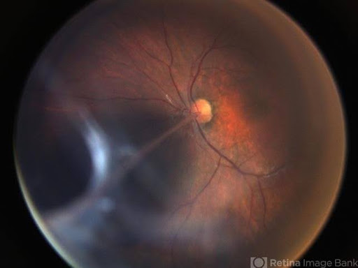
PFV is a rare condition.
It is usually diagnosed during infancy because the affected eye often appears abnormal or vision problems become apparent early in life.
Early diagnosis is important because timely treatment may improve visual outcomes and reduce complications.
Symptoms and Signs of PFV
Symptoms and signs vary depending on the severity of the condition.
Common findings include:
- Poor vision in one eye
- A white pupil reflex (leukocoria)
- Eye misalignment (strabismus)
- A small eye (microphthalmia)
- Cataract formation
- Abnormal pupil appearance
- Nystagmus (involuntary eye movements)
In infants, parents or pediatricians may notice an abnormal white reflection in photographs instead of the normal red reflex.
Types of PFV
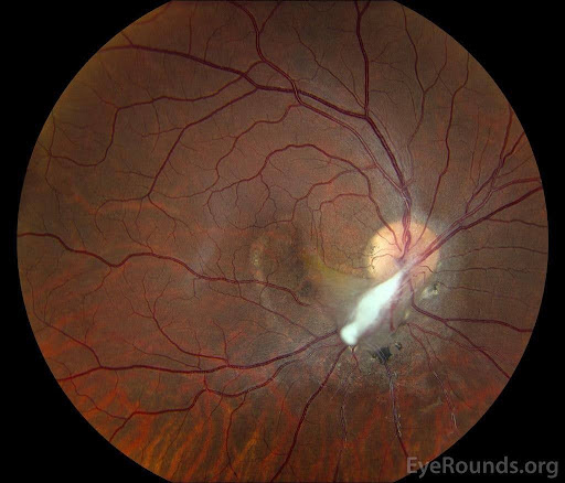
Anterior PFV
Anterior PFV primarily affects the front portion of the eye and may cause:
- Cataracts
- Elongated ciliary processes
- Shallow anterior chamber
- Lens abnormalities
Posterior PFV
Posterior PFV affects the back of the eye and may involve:
- Vitreous abnormalities
- Retinal folds
- Optic nerve abnormalities
- Retinal traction
Combined PFV
Many patients have both anterior and posterior involvement.
How Is PFV Diagnosed?

PFV is diagnosed through a comprehensive eye examination and specialized imaging.
Dilated Eye Examination
A pediatric ophthalmologist or retinal specialist evaluates the lens, vitreous, retina, and optic nerve.
Ultrasound Imaging
Ultrasound is often used to visualize internal eye structures when cataracts or other abnormalities block the view.
Optical Coherence Tomography (OCT)
OCT may help assess retinal involvement in older children who can cooperate with imaging.
Examination Under Anesthesia
Young children may require examination under anesthesia to fully evaluate the eye.
Complications of PFV
PFV can lead to a number of vision-threatening complications, including:
- Cataracts
- Glaucoma
- Retinal detachment
- Retinal folds
- Amblyopia (“lazy eye”)
- Severe vision loss
The severity of complications varies significantly from patient to patient.
Treatment for PFV

Treatment depends on the severity of the condition and the structures involved.
Observation
Very mild cases may only require monitoring.
Amblyopia Treatment
If useful vision is present, treatment may include:
- Eye patching
- Corrective glasses
- Vision therapy
Early visual rehabilitation is often important for maximizing visual development.
Surgery
Many patients require surgery to remove abnormal tissue and improve visual potential.
Surgical treatment may include:
- Cataract removal
- Vitrectomy surgery
- Release of retinal traction
- Repair of retinal detachment
The goals of surgery are to:
- Preserve the eye
- Improve vision when possible
- Reduce complications
What Is the Prognosis?

The outlook for PFV depends on:
- Severity of the disease
- Whether the retina is involved
- Timing of diagnosis
- Success of treatment
- Development of amblyopia
Mild cases may retain useful vision, while severe cases can result in significant visual impairment.
Early diagnosis and treatment generally offer the best chance for improved visual outcomes.
Living With PFV
Children with PFV often benefit from coordinated care involving:
- Pediatric ophthalmologists
- Retina specialists
- Low vision specialists
- Pediatricians
- Vision rehabilitation services
Parents should maintain regular follow-up visits and closely follow treatment recommendations to maximize visual development.
Frequently Asked Questions (FAQ)
Is PFV inherited?
Can PFV affect both eyes?
Can surgery restore normal vision?
More Information
For additional patient information, visit the American Society of Retina Specialists Persistent Fetal Vasculature Page