
Polypoidal Choroidal Vasculopathy (PCV)
Polypoidal choroidal vasculopathy (PCV) is a retinal disease in which abnormal blood vessels develop beneath the retina and form aneurysm-like enlargements called polyps. These abnormal vessels can leak fluid or blood, leading to swelling, bleeding, and vision loss.
PCV is considered a subtype of macular neovascular disease and shares many similarities with wet age-related macular degeneration (wet AMD). However, PCV has distinct characteristics and may require a different treatment approach.
Early diagnosis and treatment are important because PCV can cause recurrent bleeding and permanent central vision loss if left untreated.
What Is the Choroid?
The choroid is a layer of blood vessels located beneath the retina. It supplies oxygen and nutrients to the retina and retinal pigment epithelium (RPE).
In PCV, abnormal blood vessels develop within the choroid and beneath the retina. These vessels can form balloon-like dilations, or polyps, which are prone to leakage and bleeding.
Who Gets PCV?
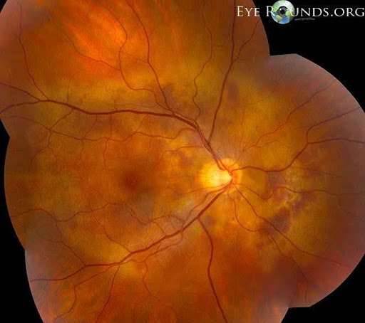
PCV is most commonly diagnosed in adults over age 50.
Risk factors may include:
- Increasing age
- High blood pressure
- Smoking
- Family history of retinal disease
PCV occurs in all ethnic groups but is more common in individuals of Asian and African ancestry than in Caucasian populations.
Many patients initially receive a diagnosis of wet macular degeneration before specialized imaging identifies PCV.
Symptoms of PCV
Symptoms depend on the amount of fluid or bleeding beneath the retina.
Common symptoms include:
- Blurred central vision
- Distorted or wavy vision
- Dark spots in central vision
- Difficulty reading
- Sudden vision loss from retinal bleeding
Symptoms often affect one eye initially but can occur in both eyes.
Some patients experience periods of stability followed by episodes of recurrent leakage or bleeding.
How Is PCV Diagnosed?
A retinal specialist diagnoses PCV using a combination of examination and advanced retinal imaging.
Optical Coherence Tomography (OCT)
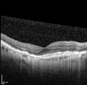
OCT provides detailed cross-sectional images of the retina and may reveal:
- Fluid beneath the retina
- Pigment epithelial detachments
- Characteristic features suggestive of PCV
Fluorescein Angiography
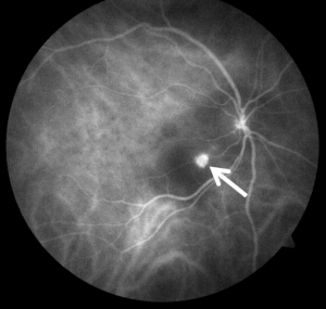
This imaging test evaluates retinal blood vessel leakage and helps assess macular damage.
Indocyanine Green Angiography (ICGA)
ICGA is considered the gold standard for diagnosing PCV.
This test uses a special dye to visualize the choroidal circulation and identify:
- Polyps
- Branching vascular networks
- Areas of leakage
Fundus Photography
Retinal photographs document bleeding, fluid, and disease progression over time.
How Is PCV Different from Wet Macular Degeneration?
PCV shares many features with wet AMD, including:
- Abnormal blood vessel growth
- Fluid leakage
- Central vision loss
However, PCV differs because:
- It forms characteristic polyp-like lesions
- It may cause larger retinal hemorrhages
- It often responds differently to treatment
- Specialized imaging is usually needed for diagnosis
Correct diagnosis is important because treatment strategies may vary.
Treatment for PCV

Modern treatments have significantly improved outcomes for patients with PCV.
Anti-VEGF Injections
Anti-VEGF medications are often the first-line treatment.
These medications help:
- Reduce fluid leakage
- Stabilize vision
- Improve retinal anatomy
Common medications include:
- Aflibercept
- Faricimab
- Ranibizumab
- Bevacizumab
Many patients require ongoing injections to maintain disease control.
Photodynamic Therapy (PDT)
Photodynamic therapy may be used in selected patients, particularly when polyps persist despite anti-VEGF treatment.
PDT involves:
- Intravenous administration of a light-sensitive medication
- Application of a specialized laser
- Closure of abnormal blood vessels
In some cases, PDT is combined with anti-VEGF therapy.
Treatment of Retinal Hemorrhage
Large retinal hemorrhages may occasionally require additional procedures or surgery.
What Is the Prognosis?
The outlook for PCV has improved dramatically with modern treatment.
Many patients:
- Maintain useful vision
- Experience reduced retinal fluid
- Achieve long-term disease control
However, PCV may be chronic and recurrent, requiring ongoing monitoring and treatment.
Untreated disease can result in:
- Permanent central vision loss
- Scarring
- Recurrent retinal bleeding
Regular follow-up with a retina specialist is essential.
Living With PCV
Patients with PCV should:
- Attend all scheduled appointments
- Monitor vision using an Amsler grid if recommended
- Report sudden visual changes immediately
- Control blood pressure
- Avoid smoking
Early treatment of recurrent disease can help preserve vision.
Frequently Asked Questions (FAQ)
Is PCV the same as wet macular degeneration?
Not exactly. PCV is considered a distinct condition with unique abnormal blood vessels, although it shares many similarities with wet AMD.
Can PCV cause blindness?
Will I need injections forever?
More Information
For additional patient information, visit the American Society of Retina Specialists Polypoidal Choroidal Vasculopathy Page