
Macular Hole
A macular hole is a small opening that develops in the macula, the central part of the retina responsible for sharp, detailed vision. The macula allows us to read, drive, recognize faces, and perform other activities that require fine visual detail.
When a macular hole forms, central vision can become blurry, distorted, or develop a dark spot. Most macular holes occur as a result of age-related changes in the vitreous, the gel-like substance that fills the inside of the eye.
Fortunately, modern retinal surgery is highly successful at repairing most macular holes and improving vision.
What Causes a Macular Hole?
The most common cause of a macular hole is age-related vitreous traction.
As we age:
- The vitreous gel gradually shrinks and becomes more liquid
- The vitreous separates from the retina
- In some cases, the vitreous pulls on the macula
- Continued traction can create a hole in the retinal tissue
Most macular holes occur in people over age 60 and are more common in women than men.
Less common causes include:
- Eye trauma
- High myopia (severe nearsightedness)
- Retinal detachment
- Diabetic eye disease
- Chronic macular swelling
Symptoms of a Macular Hole
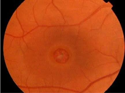
Symptoms typically affect central vision and may worsen gradually.
Common symptoms include:
- Blurred central vision
- Distorted or wavy vision
- Difficulty reading
- Difficulty recognizing faces
- A dark or missing spot in central vision
- Reduced ability to see fine details
Peripheral (side) vision usually remains normal.
Many patients first notice that straight lines appear bent or distorted.
Stages of Macular Holes
Macular holes may progress through several stages:
Impending Macular Hole (Stage 1)
At this early stage, traction affects the macula but a full-thickness hole has not yet formed.
Some stage 1 holes may resolve without surgery.
Full-Thickness Macular Hole (Stages 2–4)
A complete opening develops through the retinal layers of the macula.
As the hole enlarges, vision often becomes more significantly affected.
How Is a Macular Hole Diagnosed?
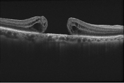
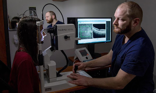
A retinal specialist can diagnose a macular hole through a dilated eye examination and specialized imaging.
Optical Coherence Tomography (OCT)
OCT is the most important test for diagnosing a macular hole.
It provides detailed cross-sectional images of the retina and allows the doctor to:
- Confirm the diagnosis
- Measure hole size
- Determine the stage
- Monitor healing after surgery
Dilated Retinal Examination
The retina specialist examines the macula and evaluates for other retinal conditions.
Amsler Grid Testing
Patients may notice distortion or missing areas when viewing an Amsler grid.
Treatment for Macular Hole
Treatment depends on the stage and severity of the hole.
Observation
Some early stage 1 macular holes may be monitored if vision remains relatively good.
However, most full-thickness macular holes require surgery.
Vitrectomy Surgery
The standard treatment for a full-thickness macular hole is pars plana vitrectomy.
During surgery:
- The vitreous gel is removed
- Traction on the macula is relieved
- A thin internal retinal layer (internal limiting membrane) is often peeled
- A gas bubble is placed inside the eye
The gas bubble helps the edges of the hole close during healing.
Positioning After Surgery
Depending on the size and type of hole, patients may be asked to maintain specific head positioning after surgery.
Your retina specialist will provide individualized instructions regarding postoperative positioning
What Is Recovery Like?
Vision improvement is usually gradual.
Patients may notice:
- Reduced distortion
- Improved reading vision
- Better central visual clarity
Recovery can continue for several months after surgery.
The amount of visual improvement depends on:
- Hole size
- Duration of the hole
- Retinal health before surgery
- Successful hole closure
Earlier treatment generally results in better outcomes.
What Is the Prognosis?
The prognosis for macular hole surgery is excellent.
Modern surgical techniques successfully close most macular holes.
Many patients experience:
- Significant visual improvement
- Better reading ability
- Reduced distortion
However, vision may not return completely to normal, especially if the hole was large or present for a long time before treatment.
Can a Macular Hole Occur in the Other Eye?
Yes. Patients who develop a macular hole in one eye have an increased risk of developing a hole in the fellow eye.
Regular monitoring is important, and patients should report any new distortion or blurred central vision promptly.
Frequently Asked Questions (FAQ)
Can a macular hole heal on its own?
Is macular hole surgery successful?
Yes. Vitrectomy surgery has a very high success rate for closing macular holes and improving vision.
Will I regain normal vision after surgery?
Many patients experience significant improvement, but the final visual outcome depends on how long the hole was present and the condition of the retina before treatment.
More Information
For additional patient information, visit the American Society of Retina Specialists Macular Hole Page